SPL Inner Ear Atlas
The SPL Inner Ear Atlas is an human ear based on CT stans from a experimental CT scanner. It was created as a joint project between the German Cancer Research Center and the SPL.
Description
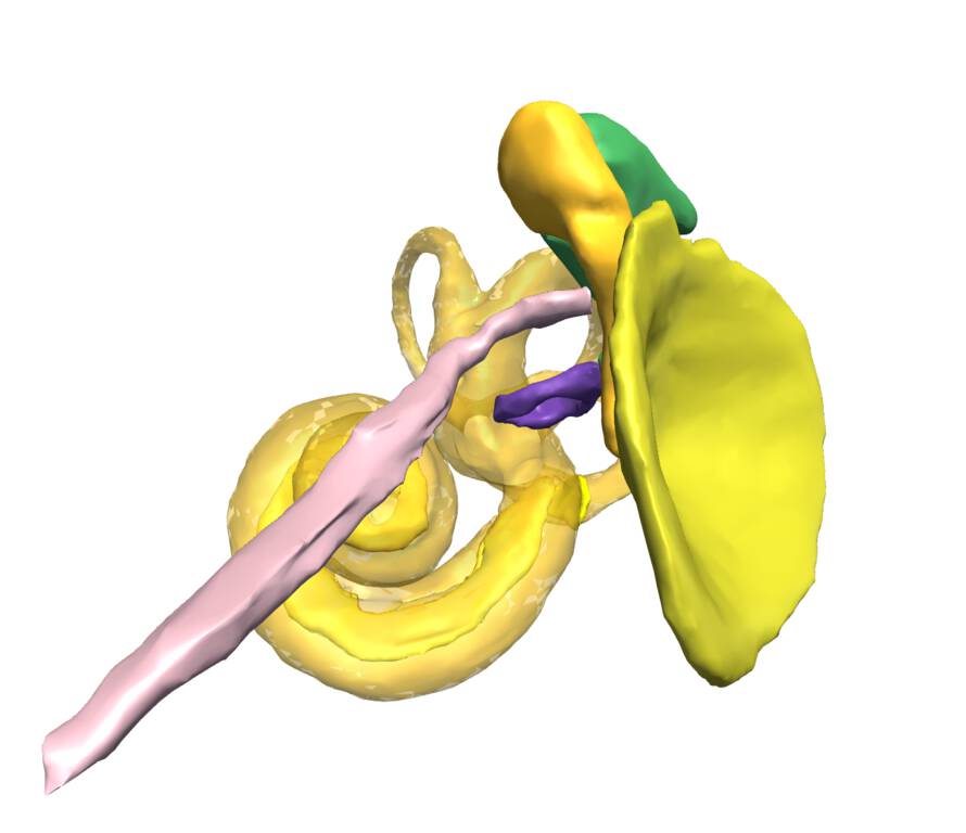
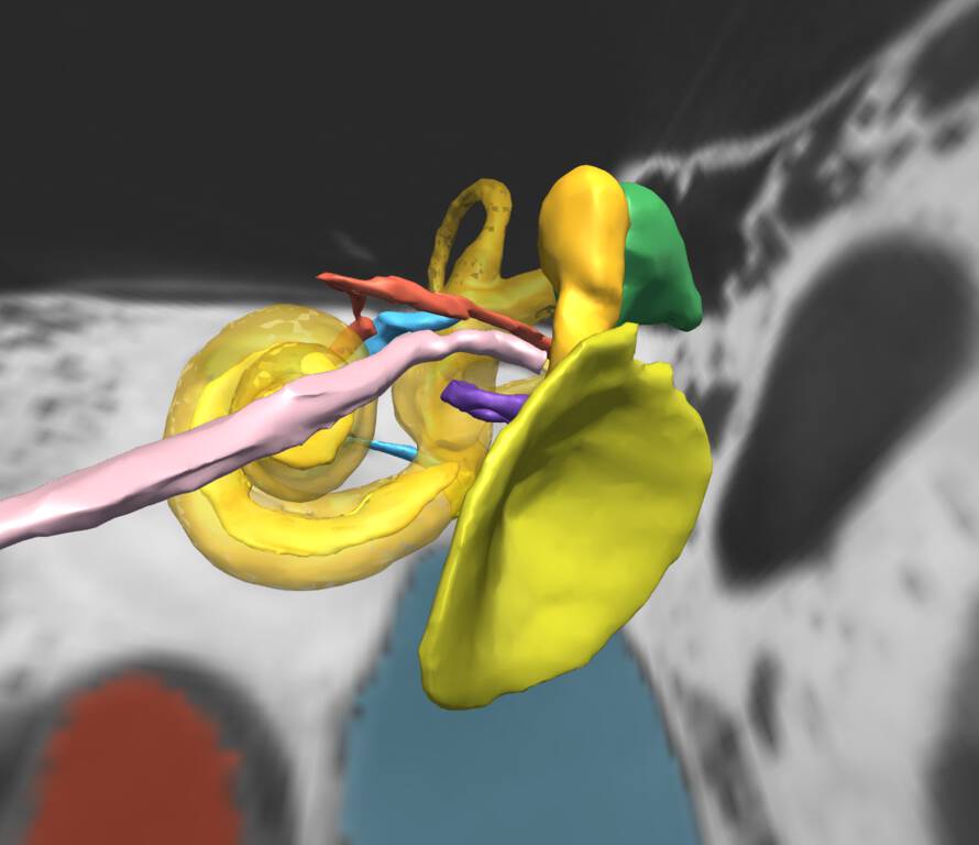
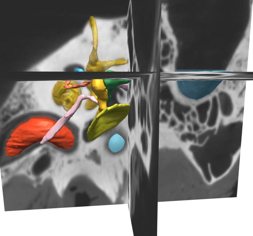
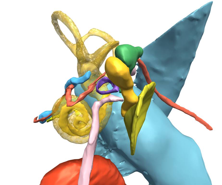
The atlas was derived from a high-resolution flat-panel computed tomography (CT) scan (aprox 140 µm high contrast resultion), using semi-automated image segmentation and three-dimensional reconstruction techniques [Gupta, Bartling, et al. AJNR Am J Neuroradiol. 2004]. The current version consists of the original CT scan; a set of detailed label maps; a set of three-dimensional models of the labeled anatomical structures.
The SPL Ear Atlas provides important reference information for surgical planning, anatomy teaching, and template driven segmentation.
Authors
- Sonke Bartling (DKFZ)
- Marianna Jakab (SPL)
- Ron Kikinis (SPL)
Institutions
- DKFZ: German Cancer Research Center, Heidelberg, Gemany.
- SPL: Surgical Planning Laboratory, Department of Radiology, Brigham and Women’s Hospital, Harvard Medical School, Boston, MA, USA.
Release date
February 2018
Keywords
atlas, ear, inner ear, CT, flat-panel CT
Sponsors and funding
- P41 RR013218/RR/NCRR NIH HHS/United States
- P41 EB015902/EB/NIBIB NIH HHS/United States
- This work is funded as part of the Neuroimaging Analysis Center, grant number P41 RR013218, by the NIH’s National Center for Research Resources (NCRR) grant number P41 EB015902, by the NIH’s National Institute of Biomedical Imaging and Bioengineering (NIBIB) and the Google Faculty Research Award.
License
Download
https://www.openanatomy.org/atlases/nac/inner-ear-2018-02.zip
References
Other images