SPL Abdominal Atlas
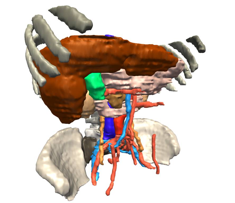
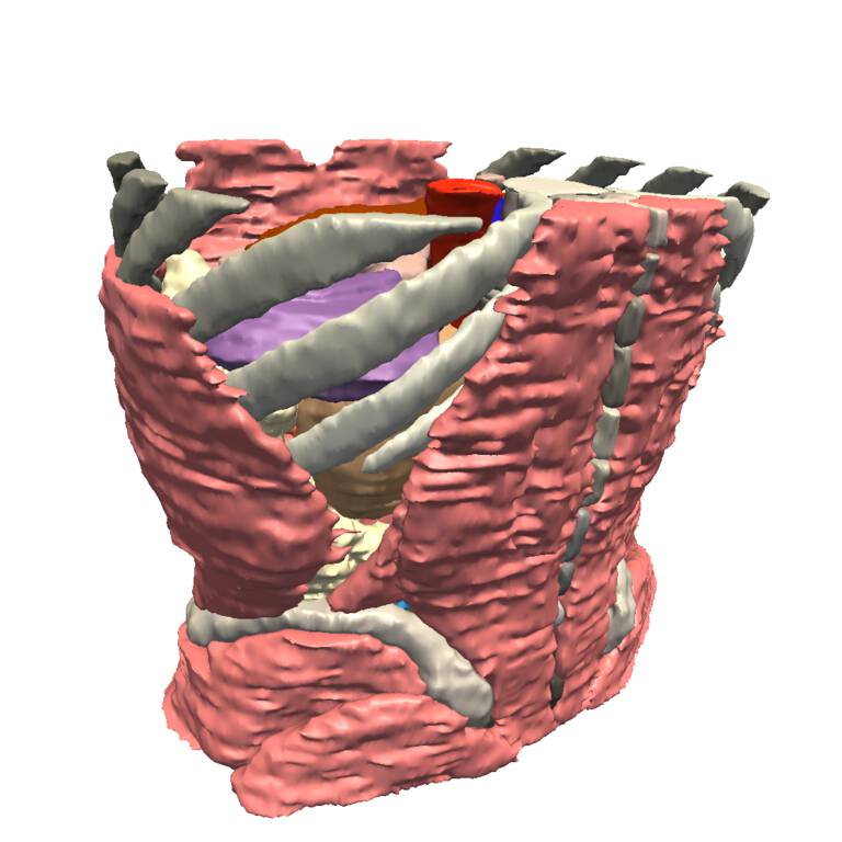
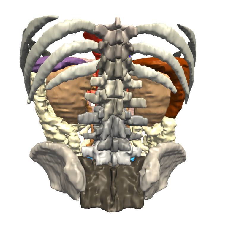
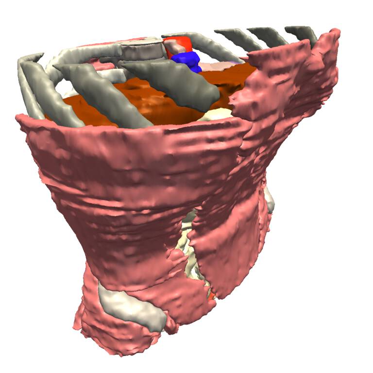
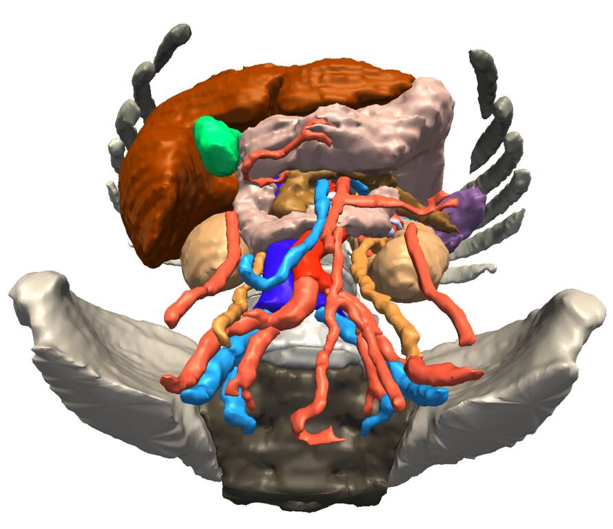
Derived from a clinical quality CT scan, this SPL Abdominal Atlas features the skeletal system, vasculature, muscles, and abdominal organs.
Description
The atlas was derived from a computed tomography (CT) scan, using semi-automated image segmentation and three-dimensional reconstruction techniques. The current version consists of the original CT scan, a set of detailed label maps, and a set of three-dimensional models of the labeled anatomical structures.
Authors
- Florin Talos (SPL)
- Marianna Jakab (SPL)
- Ron Kikinis (SPL)
Institutions
- SPL: Surgical Planning Laboratory, Department of Radiology, Brigham and Women’s Hospital, Harvard Medical School, Boston, MA, USA.
Release date
September 2015
Keywords
atlas, abdomen, CT
Sponsors and funding
- P41 RR013218/RR/NCRR NIH HHS/United States
- P41 EB015902/EB/NIBIB NIH HHS/United States
- This work is funded as part of the Neuroimaging Analysis Center, grant number P41 RR013218, by the NIH’s National Center for Research Resources (NCRR) grant number P41 EB015902, by the NIH’s National Institute of Biomedical Imaging and Bioengineering (NIBIB) and the Google Faculty Research Award.
License
Download
https://www.openanatomy.org/atlases/nac/abdomen-2016-09.zip
Other images